
Single Photon Emission Computed Tomography
Product Details:
Single Photon Emission Computed Tomography Price And Quantity
- 10 Kilograms
- 60.00 - 85.00 USD ($)/Kilograms
Single Photon Emission Computed Tomography Trade Information
- 10000 Kilograms Per Month
- 25 Days
Product Description
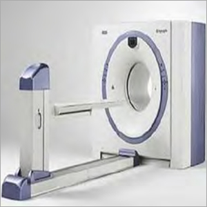
Single Photon Emission Computed Tomography (SPECT, or less commonly, SPET) is a nuclear medicine tomographic imaging technique applies gamma rays. It is quite identical to the traditional nuclear medicine planar imaging employing a Gamma Camera. However, it is able to give real 3D information. This information is basically put forth as cross-sectional slices through the patient, but can be easily re-formatted or manipulated as needed.
The technique necessitates the supply of a gamma-emitting radioisotope (a radionuclide) to the patient, normally by means of injection into the bloodstream. A marker radioisotope is added to the particular ligand to produce a radio ligand, whose features tie it to specific types of tissues. This binding allows the combination of ligand and radiopharmaceutical to be driven and confined to a place of interest in the body, where the ligand accumulation is viewed by a gamma camera.
The Applications of Tungsten Shielding in Single Photon Emission Computed Tomography
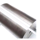
Because of its excellent density, outstanding absorption feature against radiation as well as environment-friendly attributes, tungsten alloy may extensively used in Gamma Camera (SPECT) that uses radioactive materials incorportaed as tungsten alloy syringe shield. Radiation is an efficient outfit within medicine for diagnostics as well as treatment of patients. Techniques including SPECT employ radioactive materials insinuated into the patient, which are subsequently checked by single photon emission computed tomography (SPECT) to find out the presence of tumors in the body.
Tungsten radiation shielding is a crucial device of Gamma Camera (SPECT) to fix radiation resources. Tungsten radiation shielding for SPECT can be employed together with any gamma imaging camers, where a real 3D presentation can be useful for bone scintigraphy, infection (leukocyte) imaging, thyroid imaging, tumor imaging.
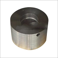
As SPECT assures of correct localization in 3D space, it can be employed to give precise information about localized function in internal organs like brain or cardiac imaging. Tungsten radiation shielding also can be applied in the syringe shield for SPECT scanner. 2 mm solid tungsten flange assists the hand when drawing out liquid from a vial. Flange is easily detached to permit the transition from giving out dose to patient injection. 9 mm thick glass-5.2g/cc ensures the best possible protection of any glass in any syringe shield and syringe shield is smoothly changed as well as makes usre that twist-turn and the syringe are held tightly.
Why Use Tungsten Shielding in Single Photon Emission Computed Tomography?
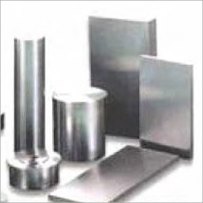
The benefits of tungsten alloy:
- High radiation reduction; excellent shielding capacity
- Thinner and often lighter than similar lead shields
- Easy to clean and sterilise
- Freely machined with traditional tools
- Strong and long-lasting no need for coating
During design of shielding, tungsten alloy radiation shielding is validated in line with needs of shield to decrease the multiple shielding materials' thickness.
Formula:
- K = e0.693 d / 1/2
- K: Shield weakened multiple
- 1/2: The tungsten alloy radiation protective material gives the half-value layer
Other Products in 'Tungsten Radiation Shield' category
- Building A2, Xinma Jingu Industrial Park, No. 959 Tianyi Road, Tianyuan District,Zhuzhou Hunan, China
- Phone :View Number
- Mr KJ Gao (CEO)
- Mobile :View Number
- Ms. Sandy Lu (Sales Executive)
- Mobile :View Number
- Send Inquiry
- International Sales Director
Mr. Jack Zhou
Mobile: +1 647 284 3766
|
Developed and Managed by Infocom Network Private Limited.


